
Application Note
Analyze cardiomyocyte spheroid contractions with SoftMax Pro Software
- Analyze numeric data from image-based experiments
- Quickly quantify beat patterns of cardiomyocytes with SoftMax Pro Peak Pro analysis algorithms
- Import raw data acquired from any scientific instrument for analysis using one software system
Introduction
Cell-based models for compound screening have become more complex in order to represent complexity of biological systems. Live-cell imaging and threedimensional models provide valuable insight into the structure and cellular processes of living cells. There have been great advances recently in developing complex organoid models representing different types of human tissues including liver1 , pancreatic2, neuronal3 , and cardiac tissues4 . Cardiac spheroids can contract in vitro and respond to the effects of different modulators. Here we describe a method that includes imaging and analysis protocols that can be used for monitoring and characterization of beating patterns of cardiac spheroids in vitro.
In the case of cardiotoxicity studies, the ability to view real-time behaviors of beating cardiomyocytes allows scientists to collect multiparametric results from a single assay through use of transmitted light, phase-contrast, and fluorescent imaging. Screening for compound cardiotoxicity during the early stages of drug development can greatly reduce costs associated with late phase drug failure due to adverse cardiovascular effects. This has led to the development of biologicallyrelevant, high-throughput, and high-content screening tools such as cardiomyocyte spheroids.
High-content screening assays produce large amounts of data with a need for rapid and automated analysis. The SoftMax® Pro Import Feature allows you to analyze numerical data from any scientific source including high-content imaging systems (Figure 1). By importing image-based fluorescence intensity data into SoftMax Pro Software, we were able to illustrate and quantify the beating patterns of cardiomyocyte spheroids treated with a panel of cardiotoxic compounds.
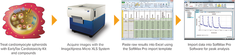
Figure 1. SoftMax Pro v6.4.2 import workflow.
Materials and methods
Spheroid rat myocardial microtissues (InSphero AG, Switzerland) were cultured in a 96-well GravityTRAP™ (InSphero AG) assay plate. Spheroids were treated with calcium dye (EarlyTox™ Cardiotoxicity Kit, #R8210, Molecular Devices) for two hours prior to addition of DMSO, 0.1 µM isoproterenol, 0.1 µM propranolol, 1 µM verapamil, 1 µM cisapride, or 1 µM lidocaine for an additional 30 minutes.
Treated spheroids were imaged at 10x magnification ten times per second in timelapse mode for a total of 300 time points using an ImageXpress® Micro XLS System, a widefield automated (1x-100x) microscope capable of fluorescent, transmitted light, and phase-contrast imaging of fixedor live-cell assays, tissues and small organisms.
Time-lapse images were quantified with MetaXpress® High-Content Image Acquisition and Analysis Software as measurements of fluorescence intensity (FI). The kinetic FI values were exported to Microsoft Excel, arranged into the Excelbased import template, and imported into SoftMax Pro v6.4.2. Data was analyzed using the pre-written Cardiomyocyte Beating i3 (Advanced) protocol, available for download at www.softmaxpro.com, to automatically calculate fourteen peak attributes: peak count, peak frequency, peak amplitude, average peak width and standard deviation, average peak rise time and standard deviation, average peak decay time and standard deviation, average peak spacing and standard deviation, average peak bottom width and standard deviation, and peak regularity.
Results
In this study, beating spheroid rat myocardial microtissues from InSphero were treated with a panel of cardiotoxic compounds and assayed with the EarlyTox Cardiotoxicity Kit containing a calcium sensitive dye optimized to measure the changes in cytoplasmic calcium concentration, which are associated with cardiomyocyte contractions. Time-lapse images of the treated cardiomyocyte spheroids were taken using the ImageXpress Micro XLS system to observe beating behavior (Figure 2). The resulting oscillations in fluorescence intensity were imported into SoftMax Pro v6.4.2 via the Excel-based import feature template to quantify peak parameters and the effects of the compounds on cardiac beating (Figure 3a). The beat frequency of isoproternoltreated cardiomyocyte spheroids increased while the beat frequency of propranolol-, cisapride-, and lidocaine-treated cardiomyocyte spheroids decreased compared to the control. In addition to decreasing beat frequency, cisapride treatment disrupted the beating pattern of cardiomyocyte spheroids. Treatment of cardiomyocyte spheroids with verapamil, a calcium channel blocker, inhibited the calcium signal (Figures 3b and 4). Taken together, the results are consistent with previous studies5,6.
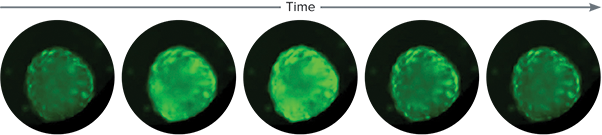
Figure 2. Cardiomyocyte spheroid time-lapse images. This image sequence shows one beat of the control rat cardiomyocyte spheroid (InSphero AG), which corresponds with an increase of cytoplasmic calcium as evidenced by an increase in fluorescent signal from the calcium sensitive dye.
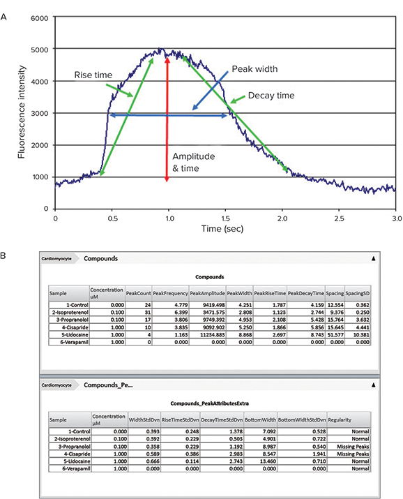
Figure 3. Peak Pro™ analysis algorithms. (A) The Peak Pro analysis algorithms allow for automated peak parameter calculation including peak frequency, count, amplitude, width (at 10% and 50% height), and spacing, rise time, decay time, and irregular spacing. (B) Display of calculated peak attributes in SoftMax Pro Software using the Cardiomyocyte Beating i3 (Advanced) protocol, available for download at www.softmaxpro.com.
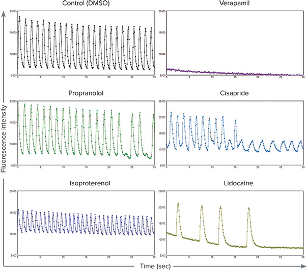
Figure 4. Cardiomyocyte spheroid beat patterns. The beat patterns of control (DMSO) and treated cardiomyocyte spheroids were visually represented in SoftMax Pro Software as plots of fluorescence intensity over a time period of 30 seconds. Top: Control (left) and verapamil (right). Middle: Propranolol (left) and cisapride (right). Bottom: Isoproterenol (left) and lidocaine (right).
Conclusions
Using biologically-relevant, cell-based models to run high-content and highthroughput cardiotoxicity screening assays that eliminate cardiotoxic drug candidates in the early stages of development could greatly reduce the time and cost of drug development. To maintain the efficiency of high-throughput and high-content screening assays, equally rapid data analysis is required. The SoftMax Pro Import Feature, available for SoftMax Pro v6.4.1 or higher, enables analysis of numerical data in microplate format acquired from a variety of scientific instruments including imaging systems, real-time PCR systems, non-Molecular Devices plate readers, scintillation counters, and more. Two import options, Excel-based and XML-based, provide a range of automation functionality from manual import to fully automated data import and analysis. In addition to the Peak Pro software algorithm and protocol, which are specifically used to quantify multi-peak oscillating data such as cardiomyocyte and cardiomyocyte spheroid beating, SoftMax Pro’s formula system and Syntax Helper help to achieve the results you need including IC50 plots and conditional pass/ fail results. Customized protocols may be saved for repeated use, ensuring efficient and consistent analysis regardless of the data source.
References
- Kermanizadeh A, Løhr M, Roursgaard M, et al. Hepatic toxicology following single and multiple exposure of engineered nanomaterials utilising a novel primary human 3D liver microtissue model. Particle and Fibre Toxicology. 2014; 11:56. li>
- Zuellig RA, Cavallari G, Gerber P, Tschopp O, Spinas GA, Moritz W, and Lehmann R. Improved physiological properties of gravity-enforced reassembled rat and human pancreatic pseudo-islets. Journal of Tissue Engineering and Regenerative Medicine. 2014 doi: 10.1002/term.1891
- Kraus D, Boyle V, Leibig N, Stark GB, and Penna V. The Neuro-spheroid—A novel 3D in vitro model for peripheral nerve regeneration. Journal of Neuroscience Methods. 2015; 246:97-105.
- Zuppinger C, Agarkova I, Moritz W, and Kelm J. A human 3D myocardial microtissue model for cardiotoxicity testing. Poster presented at: EUROTOX 2013. 49th Congress of the European Societies of Toxicology. 2013 Sep 1-4; Interlaken, Switzerland.
- Sirenko O, Cromwell EF, Crittenden C, Wignall JA, Wright FA, and Rusyn I. Assessment of beating parameters in human induced pluripotent stem cells enables quantitative in vitro screening for cardiotoxicity. Toxicology and Applied Pharmacology. 2013; 273(3):500-507.
- Sirenko O, Crittenden C, Callamaras N, Hesley J, Chen YW, Funes C, Rusyn I, Anson B, and Cromwell EF. Multiparameter In Vitro Assessment of Compound Effects on Cardiomyocyte Physiology Using iPSC Cells. Journal of Biomolecular Screening. 2013; 18:39-53.