Disease Modeling with 3D Cell-Based Assays Using a Novel Flowchip System and High-Content Imaging
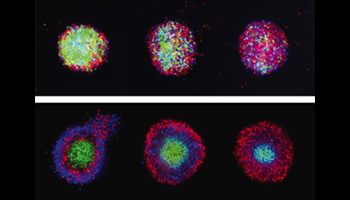
There is an increasing interest in using three-dimensional (3D) cell structures for modeling tumors, organs, and tissue to accelerate translational research. We describe here a novel automated organoid assay system (the Pu·MA System) combined with microfluidic-based flowchips that can facilitate 3D cell-based assays. The flowchip is composed of sample wells, which contain organoids, connected to additional multiple wells that can hold various assay reagents. Organoids are positioned in a protected chamber in sample wells, and fluids are exchanged from side reservoirs using pressure-driven flow. Media exchange, sample staining, wash steps, and other processes can be performed without disruption to or loss of 3D sample. The bottom of the sample chamber is thin, optically clear plastic compatible with high-content imaging (HCI). The whole system can be kept in an incubator, allowing long-term cellular assays to be performed. We present two examples of use of the system for biological research. In the first example, cytotoxicity effects of anticancer drugs were evaluated on HeLa and HepG2 spheroids using HCI and vascular endothelial growth factor expression.