Application Note
Optimized settings for Transcreener TR-FRET assays on SpectraMax iD5 and i3x readers
- Single-addition, mix-and-read assay format for high-throughput screening
- Minimal interference from fluorescent compounds with red tracer and time-resolved readout
- Z’ factor > 0.7 at 10% conversion of 10 μM ATP
Joyce Itatani | Applications Scientist | Molecular Devices
Cathy Olsen | Sr. Applications Scientist | Molecular Devices
Introduction
Transcreener® HTS is a universal, high throughput biochemical assay platform based on the detection of nucleotides, which are formed by thousands of cellular enzymes—many of which catalyze the covalent regulatory reactions that are central to cell signaling and are of great value as targets in drug discovery.
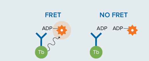
The Transcreener® TR-FRET Assays are single step, competitive immunoassays for direct detection of nucleotides with a far-red time-resolved Förster resonance energy transfer (TR-FRET) readout. The reagents for all of the assays are a far-red tracer bound to a highly specific monoclonal antibody-terbium conjugate. Excitation of the terbium complex in the UV range (ca. 330 nm) results in energy transfer to the tracer and emission at a higher wavelength (665 nm). Nucleotide diphosphate or monophosphate produced by the target enzyme displaces the tracer from the antibody, leading to a decrease in TR-FRET (Figure 1). The use of a red tracer minimizes interference from fluorescent compounds and light scattering. The Transcreener TR-FRET Assays are designed specifically for high-throughput screening (HTS) with a single addition, mix-and-read format.

Figure 1. Transcreener TR-FRET assay principle. This competitive assay uses a donor-labeled antibody and a fluorescently labeled tracer. When no analyte is present, FRET occurs. In the presence of analyte, the tracer is competed off the antibody, and FRET is disrupted.
Validation criteria
A critical factor in realizing the advantages of the Transcreener HTS assays is the correct setup of the microplate reader used for data readout. Proper selection of instrument settings, including filters and other components, can impact the instrument’s sensitivity for any given assay. The key instrument parameters for Transcreener HTS assay performance were identified by running a 10 μM ATP/ADP standard curve (24 replicates), as standard curves of this type mimic enzyme reactions. Starting with 10 μM ATP, ADP was added in increasing amounts and ATP was decreased proportionately, maintaining a total adenine nucleotide concentration of 10 μM. The flash numbers were varied to determine the requirements for a Z’ > 0.5. An instrument is validated for use with Transcreener TR-FRET assays if a Z’ > 0.7 at 10% conversion of ATP is achieved.
Materials
- Transcreener ADP 2 TR-FRET Red Assay (cat. #3011-1K)
- ATP/ADP Mixture: 10 mM MgCl 2 , 20 mM HEPES, 0.01% Brij-35 and ATP/ADP (combined to a constant adenine concentration of 10 μM)
- ADP Detection Mixture: 1X Stop and Detect Buffer C, 8 nM ADP2 Antibody-Tb Conjugate, 26.8 nM ADP HiLyte 647 Tracer
- High FRET Mixture: 8 nM ADP 2 Antibody-Tb Conjugate, 26.8 nM ADP HiLyte 647 Tracer, 1X Stop and Detect Buffer C, 10 μM ATP
- Low FRET Mixture: 8 nM ADP 2 Antibody-Tb Conjugate, 26.8 nM ADP HiLyte 647 Tracer, 1X Stop and Detect Buffer C, 10 μM ADP
Standard curve setup
- Dispense 10 μL of each ATP/ADP combination across the entire row of a 384-well plate.
- Add 10 μL of ADP Detection Mixture to those rows.
- Dispense 10 μL of the 10 μM ATP/0 uM ADP combination into row P.
- Dispense 10 μL of High FRET Mixture into wells P1-P12.
- Dispense 10 μL of Low FRET Mixture into wells P13-P24.
For a more detailed procedure on preparation of a standard curve, please refer to the appropriate Transcreener Technical Manual (https://www.bellbrooklabs.com/technical-resources/technical-manuals/).
Microplate readers
- SpectraMax® iD5 Multi-Mode Microplate Reader (Molecular Devices cat. #ID5-STD), equipped with the following:
- Enhanced TRF Module (Molecular Devices cat. #0200-7030)
- Filter 320/100 (available from Molecular Devices)
- Filter 616/10 (Molecular Devices cat. #6590-0118)
- Filter 665/10 (Molecular Devices cat. #6590-0121)
- SpectraMax i3x® Multi-Mode Microplate Reader (Molecular Devices cat. #I3X), equipped with the following:
- HTRF Detection Cartridge (Molecular Devices cat. #0200-7011)
Instrument setup
The SpectraMax iD5 and SpectraMax i3x readers use SoftMax® Pro Data Acquisition and Analysis Software. Proceed with the following steps using optimized settings.
- Open the SoftMax Pro Software and select TR-FRET for the Read Mode and Endpoint for the Read Type. On the SpectraMax i3x reader, HTRF (cartridge) should be selected as the Optical Configuration.
- On the SpectraMax iD5 reader, install the 320/100 excitation filter and 616/10 and 665/10 emission filters, and then select these filters from the Excitation and Emission dropdown wavelength menus. On the SpectraMax i3x reader, install the HTRF cartridge, and wavelengths will be selected automatically when HTRF is selected as the Optical Configuration.
- Plate Type and Read Area should be selected based on the microplate used and wells to be read. For the standard curve described, all wells of the microplate are read.
- PMT and Options Settings: Set Flashes per Read (or Pulses per Read) to 20 to 100 (for results shown here, 50 was used), measurement delay to 0.05 ms, and integration time to 0.7 ms.
- Microplate and read height should be optimized for each new lot of microplates or assay volume used.
The same instrument settings can be used to read subsequent plates as long as the volumes, microplate lot, tracer, and concentrations remain the same. Table 1 summarizes the settings described above.
Select ‘Use Filter’ for both Ex, Em.
Excitation: 320 nm
Emission 1: 615 nm
Emission 2: 665 nm
Excitation: 320 nm
Emission 1: 615 nm
Emission 2: 665 nm
Flashes per read: 50
Excitation time: 0.05 ms
Measurement delay: 0.05 ms
Integration time: 0.7 ms
Read height: 5.53 mm from the plate
On-the-fly detection: Off – Stop and Go
Number of pulses: 50
Excitation time: 0.05 ms
Measurement delay: 0.05 ms
Integration time: 0.7 ms
Read height: 6.46 mm from the plate
Table 1. Recommended settings for SpectraMax iD5 and i3x readers. Microplate and read height should be optimized for each new lot of microplates or assay volume used (use the Show Pre-Read Optimization options in the instrument settings in SoftMax Pro Software).
Conclusion
We demonstrate the validation of SpectraMax iD5 and SpectraMax i3x readers for use with the Transcreener ADP2 TR-FRET Red Assay. By using the optimized instrument settings, Z’ values > 0.8 are achievable (Table 2). For both instruments, Z’ values above the validation criterion of 0.7 were reached at ATP conversion rates of 2% or more (Figure 2), exceeding the minimum validation requirement of a Z’ greater than 0.7 at 10% ATP conversion.
Table 2. Assay performance with different reader settings

Figure 2. A) Z’ values observed in a standard curve mimic conversion of 10 μM ATP to ADP. B) Zoomed view of the 0–2 μM ADP section of the standard curve shows the Z’ validation minimal qualification data (red dotted line) and 10% ATP conversion validation point (black dotted line). Blue plots show data from the SpectraMax iD5 reader, and green plots show data from the SpectraMax i3x reader, both with 50-ms integration time.
Joyce Itatani | Applications Scientist | Molecular Devices
Cathy Olsen | Sr. Applications Scientist | Molecular Devices
介绍
Transcreener® HTS 是一种通用的,高通量的生化测定平台, 该测定法是基于核苷酸 (ADP) 的检测,核苷酸可由数千种激 酶催化形成,其中很多激酶的催化共价调节反应对细胞的信 号传导至关重要,并作为靶标在药物发现中具有重要价值。
Transcreener®TR-FRET 测定法是一步竞争 性免疫测定法, 可直接检测带有远红荧光标记的核苷酸的荧光信号。该测定 法是基于时间分辨荧光的共振能量转移 (TR-FRET) 的原理。 该测定体系中使用的是与高特异性单克隆抗体 - 淬灭剂偶合 物结合的远红示踪剂,在紫外范围 ( 约 330 nm ) 激发铽 (Tb) 配 合 物 会导致 其能 量转 移到示 踪 剂,并 在 更高的 波长 (665 nm) 产生发射光。目标酶产生的二磷酸或单磷酸核苷酸置换 了与 抗 体 链 接 的 示 踪 剂,导 致 TR-FRET 降 低 ( 图 1)。使 用 红色示踪剂可最大程度地减少荧光化合物和光散射的干扰。 Transcreener TR-FRET 测定法 是专为 HTS 设 计的,采 用一 步添加,混合和读取的测定模式。

图 1 Transcreener TR-FRET 测定原理。 该竞争性测定法使用了供体标记的抗体和作 为受体的远红示踪剂。 当不存在分析物时,会发生 FRET。 在分析物存在下,示踪剂与抗 体竞争,FRET 被破坏。
验证要求
实 现 Transcreener HTS 分 析 优 势 的 一 个 关 键 因 素 是 获 取 数 据 的 酶 标 仪 的正 确 设 置,是 否 正 确 选 择 仪器 设 置 ( 包 括 滤光片和其他组件 ) 可能会影响仪器对任何给定测定的灵敏 度。通过运行 10 μM ATP/ADP 标准曲线 (24 次重复 ) 来确定 Transcreener HTS 检测性能的关键仪器参数,并使用该标准 曲线模拟酶促反应。从 10μM 浓度的 ATP 开始, ADP 的量慢 慢增加,ATP 的量则按比例的减少,使腺嘌呤核苷酸总浓度保 持在 10 μM。调整闪光次数来实现 Z′ > 0.5 的质量要求。如 果 ATP 在 10% 的转化率下能达到 Z′ 值> 0.7,则表明该仪器 能够用于 Transcreener TR-FRET 检测。
材料
- Transcreener ADP 2 TR-FRET Red Assay (cat. #3011-1K)
- ATP/ADP 组合液 : 10 mM MgCl2, 20 mM HEPES, 0.01% Brij-35 and ATP/ADP ( 结合成恒定的腺嘌呤浓度 10 μM )
- ADP 检测混合液 : 1X Stop and Detect Buffer C, 8 nM ADP2 Antibody-Tb Conjugate, 26.8 nM ADP HiLyte 647 Tracer
- gh FRET 混 合 液 : 8 nM ADP2 Antibody-Tb Conjugate, 26.8 nM ADP HiLyte 647 Tracer, 1X Stop and Detect Buffer C, 10 μM ATP
- Low FRET 混 合 液 : 8 nM ADP2 Antibody-Tb Conjugate, 26.8 nM ADP HiLyte 647 Tracer, 1X Stop and Detect Buffer C, 10 μM ADP
标准曲线制备
- 在 384 孔 板的整 行中分配 10 μL 的每种 ATP/ADP 组合 浓 度的组合液。
- 在这些行中加入 10 μL ADP 检测混合液。
- 将 10 μL 的 10 μM ATP/0 μM ADP 组合液分配到 P 行。
- 将 10 μL 的 High FRET 混合液分配到 P1-P12 孔中。
- 将 10 μL 的 Low FRET 混合液分配到孔 P13-P24 中。
有关制备标准曲线的详细步骤,请参阅相应的 Transcreener 技 术 手 册 (https://www.bellbrooklabs.com/technicalresources/technical-manuals/)。
微孔板读板机
- SpectraMax®iD5 多功能酶标仪 (Molecular Devices cat, #ID5-STD),配备以下组件:
- TRF 增强模块 (Molecular Devices cat.#0200-7030)
- 滤光片 320/100 ( 可由 Molecular Devices 提供 )
- 滤光片 616/10 (Molecular Devices cat. #6590-0118)
- 滤光片 665/10 (Molecular Devices cat. #6590-012)
- SpectraMax®i3x 多功能酶标仪 (Molecular Devices cat, #I3x),配备以下组件:
- HTRF 检测卡盒 (Molecular Devices cat. #0200-7011)
仪器设置
SpectraMax iD5 和 SpectraMax i3x 读板机结合 SoftMax®Pro 数据采集和分析软件, 使用优化的设置继续执行以下步骤:
- 打 开 SoftMax Pro 软 件,“ 读 数 模 式“ 选 择 TR-FRET,” 读 数 类 型“ 选 择 Endpoint; 在 SpectraMax i3x 读 板 机 上,光学配置选择 HTRF ( 卡盒 )。
- 在 SpectraMax iD5 读板机上,安装 320/100 激发滤光片 以及 616/10 和 665/10 发射滤光片,然后从激发和发射下 拉波长菜单中选择这些滤光片。在 SpectraMax i3x 读板机 上,安装 HTRF 卡盒,当选择 HTRF 作为光学配置时,波长 将自动选择。
- 根据要检测的微孔板选择微孔板的“ 类型 ”,待测的微孔 板区域选择“ 读数区域 ”。
- PMT 和选项设置:将每次读取闪烁 ( 或每次读取脉冲 ) 设 置为 20 到 100 ( 本文应用程序中设置为 50 ),测量延迟设 置为 0.05 ms,积分时间设置为 0.7 ms。
- 针对每批新的微孔板或所使用的测定体积,最好优化微孔板位置和读板的高度。
只要体积,微孔板批次,示踪剂和浓度保持不变,相同的仪 器设置可用于后续板的读取。 表 1 总结了上述设置。
Select ‘Use Filter’ for both Ex, Em.
Excitation: 320 nm
Emission 1: 615 nm
Emission 2: 665 nm
Excitation: 320 nm
Emission 1: 615 nm
Emission 2: 665 nm
Flashes per read: 50
Excitation time: 0.05 ms
Measurement delay: 0.05 ms
Integration time: 0.7 ms
Read height: 5.53 mm from the plate
On-the-fly detection: Off – Stop and Go
Number of pulses: 50
Excitation time: 0.05 ms
Measurement delay: 0.05 ms
Integration time: 0.7 ms
Read height: 6.46 mm from the plate
表 1 SpectraMax iD5 和 i3x 读板机的推荐设置。 。使用新批次的微孔板或测定体积的时候,可以对微孔板位置和读 板高度进行优化( 使用 SoftMax Pro Software 仪器设置中的“Show Pre-Read Optimization Options”选项 )
结论
在本文中,我们验证了使用 SpectraMax i3x 和 iD5 多功能读板机进行 Transcreener ADP2 TR-FRET 测定过程中表现出的优越性 能,通过使用优化设置,可以达到 Z′ 值 > 0.8 ( 表 2 )。对于这两种仪器,在 2% 或更高的 ATP 转化率下都达到了高于验证标准 0.7 的 Z′ 值 ( 图 2 ),超过了在 10% ATP 转化下 Z′ 大于 0.7 的最低验证要求。
表 2 不同读板机设置下的分析性能。

图 2 A)在模拟 10μM ATP 向 ADP 转化的标准曲线中观测到的 Z′ 值。B)标准曲线在 0-2 μM ADP 区间的放大图,显示了 Z′ 检验的最小合格数据点 ( 红色虚线 )和 10% ATP 转化的检测点( 黑色虚线 )。蓝色图显示了来自 SpectraMax iD5 读板机的数据,绿色图显示了来自 SpectraMax i3x 读板 机的数据,两者的积分时间均为 50 ms。