Application Note
Advanced kinetic analysis of a bacterial growth assay
- Save time with simultaneous detection of both cell density and fluorescent protein kinetic traces using the Workflow Editor
- Simplify data report generation with preconfigured and customizable data analysis options
- Easily consolidate desired data output parameters into table or plate formats and graph as scatter plots or bar graphs
Introduction
Michel Hoogenkamp | Research Technician | Academic Centre for Dentistry Amsterdam (ACTA)
Cathleen Salomo | Field Applications Scientist | Molecular Devices
Difficult-to-kill bacteria have become a problem in hospital-acquired infections. Identifying compounds which kill these bacteria is of interest to many pharmaceutical companies. Developing and optimizing assays to screen for the efficacy of these compounds is a challenge faced by many microbiologists.
Here, we describe the setup of a bacterial growth assay using SoftMax® Pro 7.0 (or higher) Data Acquisition and Analysis Software. Both cell density and GFP signals were recorded over a time course using the Workflow Editor acquisition function. Various data transformation steps in the software are discussed, such as normalizing the GFP signal to cell density, as well as extracting growth rates or other kinetic relevant information.
Materials
- Enterococcus faecalis strain OG1RF, containing:
- Plasmid pMV158-GFP (green fluorescent protein)
- 96-well black-walled μClear microplate (Greiner Bio-One cat. #655096)
- qPCR seal (Eurogentec cat. #RT-OPSL)
- SpectraMax i3x Multi-Mode Microplate Reader (Molecular Devices cat. #i3X) with:
- SoftMax Pro 7.0 (or higher) Software
Methods
Data acquisition using the Workflow Editor
Bacterial growth data were captured using the SpectraMax® i3x Multi-Mode Microplate Reader at the Department of Preventive Dentistry at the Academic Centre for Dentistry (ACTA) in the Netherlands. Here, Enterococcus faecalis strain OG1RF containing plasmid pMV158-GFP was used at various growth conditions. GFP (green fluorescent protein) expression is heterologous, and the strain is described in the publication of Hoogenkamp et al1, where GFP was studied as a viability marker for the difficult-to-stain species E. faecalis. 150 μl of E. faecalis in PBS, and PBS as background control, were pipetted into a 96-well black-walled μclear plate. To prevent evaporation, the microplate was covered with a qPCR seal. The microplate was then placed in a SpectraMax i3x reader and incubated at 37°C for the duration of the kinetic measurement.
Using the SoftMax Pro Workflow Editor, a kinetic cycle containing an absorbance read (PlateOD600) and a fluorescence read (PlateGFPBottom) with a 5-second linear plate shake between reads was created (Figure 1). Absorbance was measured at 600 nm (OD600) to determine the bacterial growth. GFP expression, an indicator of viability, was monitored using bottom-read fluorescence detection with excitation at 485 nm (bandwidth 9 nm) and emission at 515 nm (bandwidth 15 nm). Both data traces (absorbance and fluorescence) were recorded in two independent plate sections. The kinetic cycle was set to repeat once every 15 minutes for a total of 18 hours. Optional software features allow the user to pause and resume a kinetic read, enabling the addition of reagents during the run (not used in this experiment). The resulting dual read mode kinetic data were analyzed using SoftMax Pro Software.

Figure 1. SoftMax Pro 7 Workflow Editor for a dual read mode kinetic reading. The drag and drop feature is easy to configure and enables a customized workflow to be set up quickly.
Data analysis options using the reduction settings dialogue
As an initial step of data analysis, the user must decide whether or not to define a blank. The blank well location was set using the template editor, which offers the choice of a group blank or plate blank. A plate blank is subtracted from the raw data of all sample wells in the plate at each timepoint, whereas a group blank is subtracted from associated sample wells only. In this experimental setup, buffer background traces were captured as a control for possible contamination with bacteria or other species. The buffer was not interfering with either the OD600 or the GFP measurement, therefore no subtraction was required. If media or buffer components interfere with the OD600 or fluoresce, a subtraction of the blank is recommended to retrieve the true signal values for either the absorbance or fluorescence channel. Also, this allows comparability of measurements.
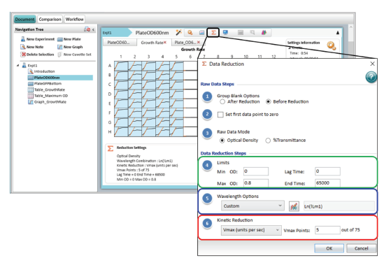
Advanced kinetic data analysis is offered through the reduction settings dialogue. Figure 2 shows the reduction settings for the bacterial growth experiment. The menu is split into two sections: raw data and data reduction steps.

Figure 2. Data analysis options in the Reduction Settings menu. This example shows the logarithmic transformation of the optical density data in plate section ‘PlateOD600nm’ and the subsequent reduction to the Vmax rate value with 5 Vmax points to retrieve the steepest part of the curve.
Raw data steps include the option to set the first data point for all kinetic traces to zero. Furthermore, a blank calculation can be included before or after the kinetic reduction. When ‘before reduction’ is selected, the averaged blank well(s) kinetic data trace is subtracted at each timepoint from all raw data sets individually. Choosing ‘after reduction’ subtracts the average of the reduced blank well data, such as Vmax rate, from the sample well data.
The data reduction steps and data output types comprise the following options (Figure 2):
- Limits: If required, limits restrict the range of kinetic data that is included in the data reduction analysis. Limits can be set for the signal-axis (OD, RLU, RFU) and/or the time-axis (seconds). The same analysis range is applied to all wells (Figure 2, green box).
- Wavelength Options: This transforms the signal-axis within the selected limits. By default, the first wavelength measured (named as !Lm1) is selected. Two examples of signal-axis transformation are shown here:
- Normalization: If the two wavelength data traces are captured within the same plate section, the dropdown menu lists preconfigured selections such as ratio (Wavelength 1/Wavelength 2 = !Lm1/!Lm2). In the example data described in this application note, the data traces were acquired in two separate plate sections, so a custom formula was required for ratiometric signal normalization. To normalize the GFP signal to the bacteria cell density, we entered a formula as follows:
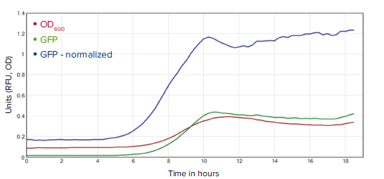
!KinPlot@PlateGFPBottom/!KinPlot@PlateOD600. This was applied in either of the two plate sections. The formula !KinPlot extracted the kinetic data set of a specified plate section referenced with the symbol ‘@’ and the corresponding name of the plate section. A comparison view of the raw data traces versus the transformed (normalized) data trace is shown in Figure 3.
- Logarithmic Scaling: To linearize the exponential growth phase of the OD
- 600
- data trace, a logarithmic transformation was applied in the OD
- 600
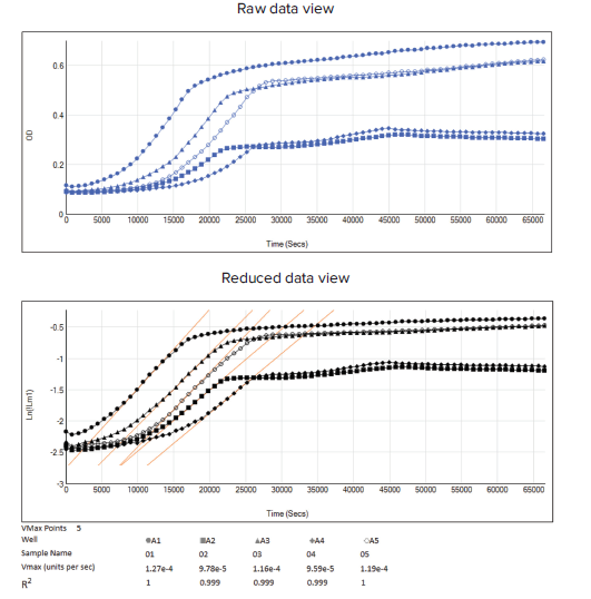
- plate section. A custom formula was added using the natural logarithm by entering Ln(!Lm1) as shown in Figure 2 (blue box). The data comparison of raw and logarithmic scaling data is shown Figure 4.
-

- Figure 4. Raw and reduced data view with logarithmic scaling applied to the OD600 data. Wells A1 to A5 are used as an example for each different experimental condition. Top: raw data of OD600 kinetic data traces (blue lines). Bottom: transformed raw data with logarithmic scaling (black lines) and the determination of the growth rate in the exponential phase by applying a Vmax data reduction (orange line). Corresponding reduction settings are shown in Figure 2.
- Normalization: If the two wavelength data traces are captured within the same plate section, the dropdown menu lists preconfigured selections such as ratio (Wavelength 1/Wavelength 2 = !Lm1/!Lm2). In the example data described in this application note, the data traces were acquired in two separate plate sections, so a custom formula was required for ratiometric signal normalization. To normalize the GFP signal to the bacteria cell density, we entered a formula as follows:
- Kinetic Reduction: This step further transforms each data trace to a reduced single value such as Vmax rate or Onset Time. A full list and details of available preconfigured kinetic reduction parameters are listed and described in Table 1. The logarithmic scaling example above is used to describe kinetic reduction:
Table 1. Preconfigured kinetic reduction options.
- As a subsequent step of the logarithmic transformation of the OD 600 data trace, the maximum growth rate can be retrieved by extracting the Vmax rate as shown in Figure 2 (red box). Vmax rate offers the advantage for users to be able to adjust Vmax points that define the maximum size of the line segment that is used to determine the slope. The Vmax is shown as an orange line in the kinetic reduced plot (Figure 4, Reduced data view). To best compare reduced kinetic graphs of all wells in the plate section view, go to Display and select ‘Reduced Data’ with the option ‘Plot’.
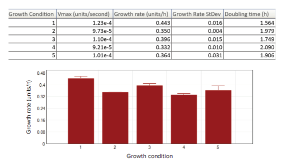
For users to best compare different growth conditions as numeric results in a tabular view, the template editor tool enables assignment of wells to sample groups, and then presents the reduced data (Vmax rate) in a table as shown in Figure 5 (upper). Through subsequent calculation steps, both growth rate (k) and doubling time (g) are obtained. For better data visualisation, the results can be shown as a bar graph (Figure 5, lower) to more easily evaluate growth conditions or treatment effects.

Figure 5. Results display options. The reduced data display of the plate section and results of each experimental condition of E. faecalis (Column 1 to 5, n=8) are summarized in the results table as wells as a bar graph. Vmax rate was used to retrieve both the growth rate (k=Vmax*3600) and doubling time (g=ln2/k).
Conclusion
The Workflow Editor in SoftMax Pro 7 Software, together with Molecular Devices multi-mode microplate readers including the SpectraMax i3x reader, offers the flexibility to record dual read mode kinetic growth data with optical density and fluorescence protein expression at the same time. Analysis is built into SoftMax Pro Software and offers a variety of data analysis options and transformation for bacterial growth data, such as data normalization or logarithmic scaling adjustment. A variety of kinetic data reduction parameters, including Vmax rate or Onset time, are preconfigured in the software. The desired data output parameters can be easily consolidated in table or plate format and graphed as bar graphs or scatter plots to support the evaluation of various experimental conditions.
Reference
- Hoogenkamp MA, Crielaard W, Krom BP. Uses and limitations of green fluorescent protein as a viability marker in Enterococcus faecalis: An observational investigation. J. Microbiol. Methods (2015) Aug;115:57-63.
前言
Michel Hoogenkamp | Research Technician | Academic Centre for Dentistry Amsterdam (ACTA)
Cathleen Salomo | Field Applications Scientist | Molecular Devices
众所周知,如何消除致病菌一直是医院获得性感染的难题。目前,许多制药公司都在研发对抗这些耐药致病菌的有效化合物,而如何筛选和鉴定这些化合物的功效是微生物学家们面临的挑战。
在本篇文章中,我们使用 SoftMax®Pro7.0 ( 或更高版本 ) 数据采集和分析软件进行细 菌生长曲线的检测。在连续的一段时间内, 使用多任务流程编辑器可以同时采集细胞 密度值和 GFP 荧光信号值。我们探讨了软 件中各种数据转换模式,例如将 GFP 信号 归一化为细胞密度,以及获取生长速率或 其他动力学相关信息。
材料
- 含有质粒 pMV 158 - GFP 的粪肠球菌菌株 OG1RF
- 黑色 96 孔底透微孔板 ( Greiner Bio - One 公司,# 655096 )
- qPCR 密封膜 ( Eurogentec 公司,#RT - OPSL )
- SpectraMax i3x 多功能微孔板读板机 ( Molecular Devices公司 ),包括SoftMax Pro 7.0 ( 或更高版本 ) 软件
方法
使用多任务流程编辑器采集数据
本篇文章中细菌生长的实验数据来自于 荷兰牙科学术中心 (ACTA) 预防牙科的 SpectraMax®i3x 多功能微孔板读板机。 在本次实验中,我们将含有质粒 pMV158 - 优势 多任务流程编辑器,可同时检 测细胞密度和荧光蛋白动态表 达,节省时间; 软件内置动力学分析参数和自 定义函数,简化数据分析流程; 轻松将数据以板格式或列表格 式输出,并绘制散点图或条形 图直观展示数据结果。 GFP的粪肠球菌菌株 OG 1 RF 作为样品并且 设置了 5 种不同的实验条件。GFP ( 绿色荧 光蛋白 ) 表达是异源的,2015 年 Hoogenkamp 等人1 报道过 GFP 是可以作为粪肠 球菌的活力标志物的。将 150 μl 的粪肠球 菌样品和空白对照 PBS 依次加入黑色 96 孔底透微孔板中,为了防止液体蒸发,最 好用 qPCR 密封膜覆盖微孔板。把微孔板 放入 SpectraMax®i3x 多功能微孔板读板机 中,设置孵育温度为 37℃,然后进行连续 时间的动力学检测。
在 SoftMax Pro 软件多任务流程编辑器 中,创建一个包含吸光度检测 (OD 600) 和 荧光强度检测 ( 荧光底读 ) 的动力学循环程 序,两次读数之间设置 5 秒钟的线性混匀 ( 见图 1 )。600 nm (OD 600) 读取的吸光度 值代表了细菌生长的密度,荧光底读所读 取的 GFP 蛋白表达的荧光强度值代表了细 菌活力,激发光设置在 485 nm ( 带宽 9 nm ),发射光设置在 515 nm ( 带宽 15 nm )。 吸光度值和荧光强度值这两种数据分别 单 独记录在吸光度检测和荧光强度检测 的程序中。此次动力学循环程序设定为每 15 分钟重复一次读数,一共持续 18 个小 时。在 SoftMax Pro 7.0 ( 或更高版本 ) 软 件中,允许暂停或恢复动力学读数,从而 便于用户在程序运行期间向微孔板中添加 试剂 ( 本次实验没有使用该功能 )。最后, 使用 SoftMax Pro 软件分析所得的双检测 模式动力学数据。

图 1 SoftMax Pro 7 多任务流程编辑器,用于双读取模式动态读数。 鼠标拖放功能易于设置,可以快速设定用户自定义的实验流程
利用数据运算功能进行数据分析
作为实验数据处理的第一步,用户必须决 定是否需要减去空白。首先,在孔板编辑 器中,定义空白孔的位置,有板空白和组 空白两种选择,板空白是指整个微孔板只 有一个空白对照,在每个时间点从板中所 有样品孔的原始数据中减去板空白;而当 对样品进行不同实验条件的分组,每组样 品设置一个组内空白,仅从相对应组别的 样品孔的原始数据中减去组空白。在本次 实验中,采集缓冲液的背景值作为是否污 染细菌或其它物质的对照。PBS 缓冲液不 干扰 OD 600 或 GFP 测量,因此不需要减 去。如果介质或缓冲液成分干扰 OD 600 或 发出荧光,建议减去空白以测定吸光度或 荧光通道的真实信号值。
通过数据处理菜单可设置先进的动态学数 据分析。图 2 展示了细菌生长实验的数据 分析设置,菜单分为两部分:原始数据设 置和数据分析设置原始数据设置中包含了 将动力学曲线的第一个数据点设置为零的 选项。此外,减去空白的操作可以在动力 学数据处理之前也可以在其之后。当选择 了“处理前”选项,将会在每个时间点从 所有样品孔的原始数据中减去空白孔的平 均值;当选择了“处理后”选项,将从样 品孔处理过的数据中再减去处理过后的空 白孔平均值。

图 2 数据处理菜单中的数据分析选项。 。该示例展示了 OD 600 nm 的吸光度数据的对数转换,并将 Vmax 值降低到 5 个 Vmax 点,以测定曲线的最陡部分。
数据分析设置和数据输出类型包括以下选 项 ( 图 2 ):
- 限值:如果需要,限值会限定数据分析中 动力学数据的范围,它可以为信号轴 (OD, RLU,RFU) 和/或时间轴 ( 秒 ) 限定范围, 对所有孔应用相同的分析范围 ( 图 2,绿色 框 )。
- 波长选项:在选定的限值范围内转换信号 轴。默认选择测量的第一个波长 ( 命名为 ! Lm 1 )。下面列举两个信号轴转换的例子: (1) 标准化:如果在同一个检测程序内读取 了两条波长的数据,则波长选项下拉菜单 会列出预置的选项,例如比率 ( 波长 1 /波长 2 =!Lm 1 /!Lm 2)。
- 在本次实验的示例数据中,两组数据来自 于两个独立的检测程序,因此比率信号归 一化需要自定义公式。为了将 GFP 荧光信 号标准化为细菌细胞密度,我们需要输入 如下公式:
!KinPlot @ PlateGFPBottom /!KinPlot @ PlateOD600。可将此公式应用于两个检 测程序中的任一个。公式!KinPlot 提取了 用符号'@'引用的指定检测程序的动力学 数据集以及相应的检测程序名称。原始数 据与转换 ( 标准化 ) 数据的比较如图 3 所 示。
- (2) 对数标度:为了使 OD 600 数据在 指数增长阶段线性化,在吸光度检测程序 中应用对数转换。通过输入 Ln (!Lm 1) 使 用自然对数添加自定义公式,如图 2 所示 ( 蓝色框 )。原始数据与对数标度数据比较 如图 4 所示。动力学分析:此选项进一步 将每个动力学数据转变成一个简化的值, 如 Vmax 最大斜率和开始时间。表 1 中是 软件内置的动力学分析参数的完整列表和 详细信息。
-

- 图 4 OD 600 原始数据曲线和转换过的对数标度曲线。 A1 孔至 A5 孔设置了 5 种不同的实验条件。 上图:OD 600 的动态原始数据曲线 ( 蓝线 ),下图:对数标度数据曲线 ( 黑线 ),并通过设置 Vmax 动态数据分析 ( 橙色线 ) 确定指数阶段的生长速率。相应的动态数据分析设置如图 2 所示。
- 在本次实验的示例数据中,两组数据来自 于两个独立的检测程序,因此比率信号归 一化需要自定义公式。为了将 GFP 荧光信 号标准化为细菌细胞密度,我们需要输入 如下公式:
- 以上面提到的对数标度转换为例来详细说明动力学分析方法:
表 1 软件内置的动力学分析参数
- OD 600 原始数据转换成对数标度数据之 后,可以通过设置 Vmax 来检测最大增长 率,如图 2 所示 ( 红色框 )。Vmax 的内置 分析选项方便了用户调整 Vmax 点,这些 点定义了用于能确定斜率的线段的最大尺 寸。Vmax 在对数标度曲线的图中显示为橙 色线 ( 图 4,对数标度曲线图例 )。另外, 在显示界面中处理数据选项里选择 “Plot”,用户可以更好的比较板视图中 所 有 孔 的 动 力 学 分 析 数 据 。 我 们 在 SoftMax Pro 7 软件孔板编辑器中将 5 种 不同实验条件的样品进行分组 (A1 - A5),
这样用户能够更直观的比较这些不同实验 组之间的数据,如图 5 ( 上图 ) 所示,表格 中显示了 Vmax 的数据,通过运算分析, 还得到了生长速率 (K) 和倍增时间 (g)。数 据结果可以显示为柱状图 ( 图 5,下图 ), 以便更直观的评估细菌的生长条件或药物 的治疗效果。

图 5 结果显示选项,选择显示分析过的数据结果。粪 。粪肠球菌每个实验组的数据结果( A1-A5列,n = 8 ) 显示在结果列表以及柱状图中。Vmax 可用于生长速率 (k = Vmax * 3600) 和倍增时间 (g = ln2 / k) 的 运算。
结论
SoftMax Pro 7 多任务流程编辑器配合 Molecular Devices SpectraMax i3x 多功 能读板机一起使用,可同时读取吸光度值 和荧光强度值,灵活的进行双检测模式的 细菌生长曲线动力学实验。在 SoftMax Pro 7 软件中,内置了各种数据分析选项和细菌 生长数据的转换分析,如吸光度和荧光强 度的标准化或对数标度调整等。软件中还 预设了各种动力学分析的简化参数,包括 最大斜率或起始时间等。动态数据输出可 以选择板格式或者列表格式输出,还可将 数据绘制成更直观的条形图或者散点图, 方便用户对不同实验条件的最终分析结果
参考文献
- Hoogenkamp MA, Crielaard W, Krom BP. Uses and limitations of green fluorescent protein as a viability marker in Enterococcus faecalis: An observational investigation. J. Microbiol. Methods (2015) Aug;115:57-63.